Home > Popular Themes > Human Body
Brain fibres, DTI MRI scan C017 / 7100

![]()
Wall Art and Photo Gifts from Science Photo Library
Brain fibres, DTI MRI scan C017 / 7100
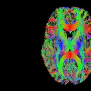
Brain fibres. 3D diffusion tensor imaging (DTI) magnetic resonance imaging (MRI) scan of a selection of nerve pathways (blue, pink, green) in the brain. The front of the brain is at left. The ventricles are yellow and the brains cortical surface is also shown. The fibres in the right hemisphere (upper left) are part of the motor system. In the left hemisphere (lower right) are functionally active areas and connections (pink) that go though them. Diffusion tensor imaging measures the direction of water diffusion, which in the brain reveals the orientation of nerve fibres. The technique is also known as tractography, with the resulting image known as a tractogram
Science Photo Library features Science and Medical images including photos and illustrations
Media ID 9268431
© SHERBROOKE CONNECTIVITY IMAGING LAB/SCIENCE PHOTO LIBRARY
Brain Imaging Brain Scan Central Nervous System Cerebral Cerebrum Diffusion Tensor Imaging Dti Scan Fiber Fibers Fibre Fibres Imaging Technique Magnetic Resonance Imaging Mri Scan Mri Scanner Nerve Nerve Fibre Nerves Neural Pathway Neural Tract Paths Pathway Pathways Structural Tractogram Tractography Ventricle Ventricles White Matter Brain Neurological Neurology
EDITORS COMMENTS
This print showcases the intricate network of brain fibres, captured through a cutting-edge imaging technique known as diffusion tensor imaging (DTI) magnetic resonance imaging (MRI). The image reveals a mesmerizing array of nerve pathways in vibrant shades of blue, pink, and green against a striking black background. Positioned at the left side is the front of the brain, while yellow ventricles and the cortical surface are also visible. In the upper left hemisphere, we observe fibres belonging to the motor system, highlighting their crucial role in coordinating movement. On the lower right hemisphere, functionally active areas and connections are depicted in pink hues. DTI measures water diffusion within the brain to unveil nerve fibre orientations accurately. This technique is commonly referred to as tractography due to its ability to map out neural tracts with exceptional precision. The resulting image is known as a tractogram – an awe-inspiring representation of our complex neurological architecture. This remarkable photograph not only offers insights into human anatomy but also underscores its significance for medicine and biology. It serves as a testament to advancements in neurology and medical imaging techniques that enable us to explore and understand our central nervous system better. Captured by Sherbrooke Connectivity Imaging Lab/Science Photo Library, this print symbolizes both beauty and scientific progress—a visual reminder of how far we have come in unraveling the mysteries hidden within our own minds.
MADE IN THE USA
Safe Shipping with 30 Day Money Back Guarantee
FREE PERSONALISATION*
We are proud to offer a range of customisation features including Personalised Captions, Color Filters and Picture Zoom Tools
SECURE PAYMENTS
We happily accept a wide range of payment options so you can pay for the things you need in the way that is most convenient for you
* Options may vary by product and licensing agreement. Zoomed Pictures can be adjusted in the Cart.