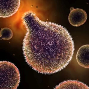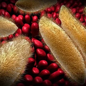
Trachea lining, TEM

![]()
Wall Art and Photo Gifts from Science Photo Library
Trachea lining, TEM
Trachea lining. Coloured transmission electron micrograph (TEM) of a transverse section through the lining of the trachea (windpipe). The trachea links the larynx to the lungs. The epithelial lining consists of cells that have hair-like cilia (turquoise) on their surface and mucus-secreting glands. Mucus (brown) secreted by the glands traps debris, such as dust particles or bacteria, in the inhaled air, while the beating of the cilia moves the mucus and particles upwards out of the respiratory tract, helping to keep the lungs and airways clear and prevent infection
Science Photo Library features Science and Medical images including photos and illustrations
Media ID 6450419
© STEVE GSCHMEISSNER/SCIENCE PHOTO LIBRARY
Cilia Cilium Glands Lining Magnified Image Microscopic Subjects Microvilli Microvillus Mucous Cell Mucus Respiratory System Secretory Trachea Transmission Electron Microscope Transverse Section False Coloured Section
EDITORS COMMENTS
This stunning coloured transmission electron micrograph (TEM) captures the intricate beauty of the trachea lining, offering a glimpse into the fascinating world within our respiratory system. The trachea, commonly known as the windpipe, serves as a vital conduit connecting the larynx to our lungs. In this image, we observe the epithelial lining composed of specialized cells adorned with hair-like cilia in a mesmerizing turquoise hue. These cilia play an essential role in maintaining our respiratory health by constantly beating and creating coordinated movements. As they sway rhythmically, they propel mucus secreted by glands (depicted in brown) upwards through our airways. The purpose of this ingenious mechanism is two-fold: first, it traps unwanted debris such as dust particles or harmful bacteria present in inhaled air; secondly, it ensures that these captured substances are effectively expelled from our respiratory tract. By keeping our lungs and airways clear from potential threats, this process helps prevent infections and maintain optimal lung function. Zooming further into this microscopic marvel reveals an array of tiny structures called microvilli on each cilium's surface. These microvilli enhance their efficiency by increasing their surface area for capturing foreign particles and facilitating mucus movement. As we admire this magnified image taken using a transmission electron microscope (TEM), we gain deeper appreciation for the intricacies hidden within us - reminding us once again how remarkable and complex our own biology truly is.
MADE IN THE USA
Safe Shipping with 30 Day Money Back Guarantee
FREE PERSONALISATION*
We are proud to offer a range of customisation features including Personalised Captions, Color Filters and Picture Zoom Tools
SECURE PAYMENTS
We happily accept a wide range of payment options so you can pay for the things you need in the way that is most convenient for you
* Options may vary by product and licensing agreement. Zoomed Pictures can be adjusted in the Cart.