Home > Animals > Mammals > Muridae > Water Mouse
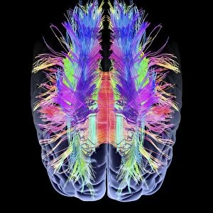
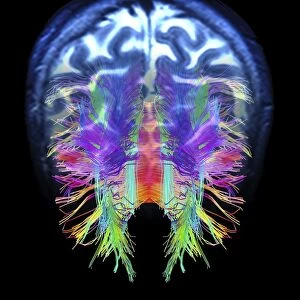
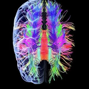
White matter fibres and brain, artwork C015 / 1932

![]()
Wall Art and Photo Gifts from Science Photo Library
White matter fibres and brain, artwork C015 / 1932
White matter fibres overlaid a 3d model of the human brain in top view. Coloured 3D diffusion spectral imaging (DSI) scan of the bundles of white matter nerve fibres in the brain. The fibres transmit nerve signals between brain regions and between the brain and the spinal cord. Diffusion spectrum imaging (DSI) is a variant of magnetic resonance imaging (MRI) in which a magnetic field maps the water contained in neuron fibers, thus mapping their criss-crossing patterns. A similar technique called diffusion tensor imaging (DTI) is also used to explore neural data of white matter fibres in the brain. Both methods allow mapping of their orientations and the connections between brain regions. Data/software: NIH Human Connectome Project /humanconnectomeproject.org)
Science Photo Library features Science and Medical images including photos and illustrations
Media ID 9211719
© PASIEKA/SCIENCE PHOTO LIBRARY
Connections Diffusion Spectral Imaging Diffusion Tensor Imaging Dsi Scan Dti Scan Fibers Fibre Tracking Fibres Human Brain Mapping Nerve Bundles Nerve Fibre Pathway Pathways White Matter Brain Connexions Nervous System
FEATURES IN THESE COLLECTIONS
> Animals
> Mammals
> Muridae
> Water Mouse
> Maps and Charts
> Related Images
EDITORS COMMENTS
This print titled "White matter fibres and brain, artwork C015 / 1932" showcases the intricate beauty of the human brain's white matter fibres. In this top view composition, a 3D model of the brain is overlaid with coloured 3D diffusion spectral imaging (DSI) scan data. The image reveals bundles of white matter nerve fibres that serve as vital pathways transmitting nerve signals between different regions of the brain and even to the spinal cord. By utilizing diffusion spectrum imaging (DSI), a variant of magnetic resonance imaging (MRI), these fibres' criss-crossing patterns are mapped by tracking water within neuron fibers using a magnetic field. Similar techniques like diffusion tensor imaging (DTI) are also employed to explore neural data related to white matter fibers in the brain. Both methods enable researchers to map orientations and connections between various areas within our complex organ. This remarkable visual representation is made possible through collaboration with NIH Human Connectome Project, an initiative dedicated to mapping human brain connectivity. The pathway-like appearance created by cutouts emphasizes how these nerve bundles form intricate networks throughout our nervous system. By showcasing this stunning artwork, we gain insight into the complexity and interconnectedness that lies within our own minds, reminding us once again of the awe-inspiring wonders found in science and nature.
MADE IN THE USA
Safe Shipping with 30 Day Money Back Guarantee
FREE PERSONALISATION*
We are proud to offer a range of customisation features including Personalised Captions, Color Filters and Picture Zoom Tools
SECURE PAYMENTS
We happily accept a wide range of payment options so you can pay for the things you need in the way that is most convenient for you
* Options may vary by product and licensing agreement. Zoomed Pictures can be adjusted in the Cart.