Left shoulder nerve plexus, artwork C016 / 6814

![]()
Wall Art and Photo Gifts from Science Photo Library
Left shoulder nerve plexus, artwork C016 / 6814
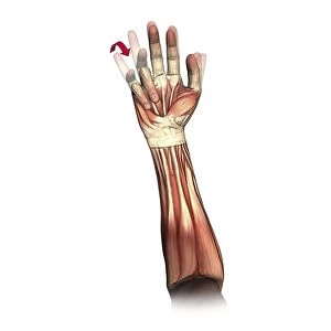
Left shoulder nerve plexus. Artwork of the nerves (yellow), arteries (red), muscles (red-brown) and bones (light brown) of the left shoulder, seen from the front. This nerve plexus lies between the clavicle bone (bottom) and the carotid artery (red). The nerve running from left is the phrenic nerve. The muscles are the infrahyoid (upper left), the sternocleidomastoid (top right), and the trapezius (centre right). The muscles of the nerve plexus are (left to right): the anterior scalene, middle scalene, and posterior scalene. For the right shoulder, see C016/6815
Science Photo Library features Science and Medical images including photos and illustrations
Media ID 9243427
© D & L GRAPHICS / SCIENCE PHOTO LIBRARY
Anterior Arteries Blood Vessels Bones Bundle Carotid Artery Clavicle Frontal Joint Muscles Nerve Nerve Plexus Nerves Neural Shoulder Sternocleidomastoid Trapezius Artery Blood Vessel Left Shoulder Neurological Neurology
EDITORS COMMENTS
This print showcases the intricate network of nerves, arteries, muscles, and bones that make up the left shoulder nerve plexus. The artwork beautifully depicts these anatomical structures in vivid detail. In this frontal view, we can observe the yellow nerves intertwining with the red arteries, while the muscles are depicted in shades of red-brown. Light brown hues represent the underlying bones that provide support and structure to this complex system. The left shoulder nerve plexus is situated between the clavicle bone at its base and the prominent carotid artery in a striking shade of red. One particular nerve stands out as it runs from left to right - known as the phrenic nerve. Highlighted within this artistic representation are three key muscles: infrahyoid (upper left), sternocleidomastoid (top right), and trapezius (centre right). These muscles play essential roles in movement and stability within the shoulder region. From left to right along this nerve plexus lie three scalene muscles: anterior scalene, middle scalene, and posterior scalene. Their positioning contributes to maintaining proper posture and facilitating various upper body movements. This visually stunning print not only serves as an educational tool for students studying anatomy but also captivates anyone interested in exploring our remarkable human biology.
MADE IN THE USA
Safe Shipping with 30 Day Money Back Guarantee
FREE PERSONALISATION*
We are proud to offer a range of customisation features including Personalised Captions, Color Filters and Picture Zoom Tools
SECURE PAYMENTS
We happily accept a wide range of payment options so you can pay for the things you need in the way that is most convenient for you
* Options may vary by product and licensing agreement. Zoomed Pictures can be adjusted in the Cart.