Home > Arts > Artists > J > Jacob Jacobs
Brain meninges

![]()
Wall Art and Photo Gifts from Science Photo Library
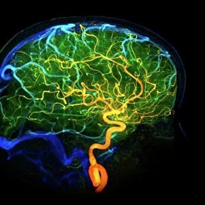
Brain meninges
Brain meninges, historical anatomical artwork. This cranial (top) view of the brain (with front at the top) and its surrounding protective meninges, which have been partially dissected away. The meninges, which cover the brain and spinal cord, consist of three layers: The dura mater (the tough, inflexible outer layer), the arachnoid mater (the delicate middle layer), and the pia mater (the fine, innermost layer). The blood vessels can be seen on the surface of the brain, with arteries red and veins and sinuses blue. This illustration is taken from the 19th century French textbook The Atlas of Human Anatomy and Surgery by J. M. Bourgery and N. H. Jacob
Science Photo Library features Science and Medical images including photos and illustrations
Media ID 6448335
© MEHAU KULYK/SCIENCE PHOTO LIBRARY
Atlas Of Human Anatomy Central Nervous System Cerebral Cranial Dissected Dissection Dura Mater French J M Bourgery Membrane Membranes Meninges N H Jacob Pia Mater Surgery Surgical Brain Nervous System Neurological Neurology
FEATURES IN THESE COLLECTIONS
> Arts
> Artists
> J
> Jacob Jacobs
EDITORS COMMENTS
This print showcases a historical anatomical artwork of the brain meninges. In this cranial view, we can observe the intricate layers of protective membranes surrounding the brain. The meninges, consisting of three distinct layers - dura mater, arachnoid mater, and pia mater - play a crucial role in safeguarding the delicate neural tissue. The image offers a glimpse into the 19th century French textbook "The Atlas of Human Anatomy and Surgery" by J. M. Bourgery and N. H. Jacob, highlighting their meticulous dissection techniques. As we examine the partially dissected brain, it becomes evident how these skilled anatomists meticulously removed sections to reveal the underlying structures. Notably visible on the surface are blood vessels that supply vital nutrients to the brain. Arteries are depicted in vivid red hues while veins and sinuses appear as calming blues. This remarkable illustration serves as a testament to both artistry and scientific exploration within biology and anatomy during that era. It provides valuable insights into our understanding of neurology, specifically focusing on cerebral anatomy and its connection with the central nervous system (CNS). As we delve into this mesmerizing piece from Science Photo Library's collection, we gain an appreciation for centuries-old medical knowledge that continues to shape our comprehension of human physiology today.
MADE IN THE USA
Safe Shipping with 30 Day Money Back Guarantee
FREE PERSONALISATION*
We are proud to offer a range of customisation features including Personalised Captions, Color Filters and Picture Zoom Tools
SECURE PAYMENTS
We happily accept a wide range of payment options so you can pay for the things you need in the way that is most convenient for you
* Options may vary by product and licensing agreement. Zoomed Pictures can be adjusted in the Cart.