Home > Animals > Mammals > Nesomyidae > Fat Mouse
Cartilage cell, TEM C014 / 1433

![]()
Wall Art and Photo Gifts from Science Photo Library
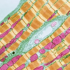
Cartilage cell, TEM C014 / 1433
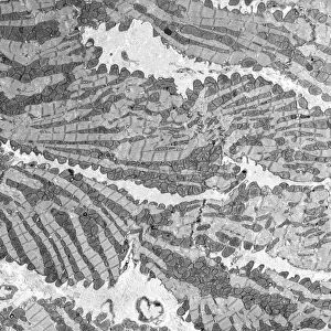
Cartilage cell. Transmission electron micrograph (TEM) of a section through a chondrocyte cell from hyaline cartilage of the trachea (windpipe). The cell nucleus (green) can be seen along with a large fat droplet (purple) and glycogen granules (bright blue). Chondrocytes are the only cells found in cartilage. They produce and maintain the cartilages extracellular matrix and. Hyaline cartilage is found in the epiphyseal plates of growing bones, and in the nose, larynx (voice box), trachea (windpipe) and bronchus. Magnification: x4000 when printed 10 centimetres wide
Science Photo Library features Science and Medical images including photos and illustrations
Media ID 9226881
© STEVE GSCHMEISSNER/SCIENCE PHOTO LIBRARY
Cartilage Cell Biology Chondrocyte Connective Tissue Cytological Cytology Extracellular Matrix Granules Histological Histology Hyaline Nucleus Trachea Transmission Electron Micrograph Transmission Electron Microscope Windpipe Section Sectioned
EDITORS COMMENTS
This print showcases the intricate details of a cartilage cell, specifically a chondrocyte from hyaline cartilage found in the trachea. Through the lens of a transmission electron microscope (TEM), we are granted an up-close view of this vital cellular component. The green nucleus stands out prominently amidst its surroundings, providing insight into the cell's genetic information and regulatory functions. What is truly fascinating about this image is the presence of other elements within the chondrocyte. A large fat droplet, depicted in purple, hints at potential energy storage or metabolic processes occurring within the cell. Additionally, bright blue glycogen granules can be observed, suggesting their role in providing fuel for cellular activities. Chondrocytes play an essential role as they are responsible for producing and maintaining the extracellular matrix that forms cartilage tissue. This connective tissue can be found not only in our trachea but also in various parts of our body such as epiphyseal plates of growing bones, nose, larynx (voice box), and bronchus. The level of magnification used to capture this image is remarkable - x4000 when printed 10 centimeters wide - allowing us to appreciate even minute details with great clarity. This photograph serves as a testament to both scientific advancement and artistic beauty by combining biology and anatomy into one visually stunning composition.
MADE IN THE USA
Safe Shipping with 30 Day Money Back Guarantee
FREE PERSONALISATION*
We are proud to offer a range of customisation features including Personalised Captions, Color Filters and Picture Zoom Tools
SECURE PAYMENTS
We happily accept a wide range of payment options so you can pay for the things you need in the way that is most convenient for you
* Options may vary by product and licensing agreement. Zoomed Pictures can be adjusted in the Cart.