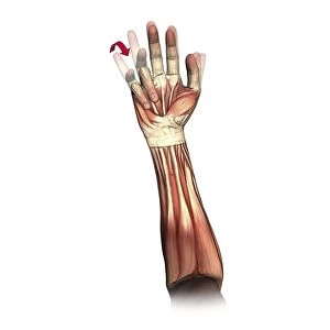
Little and ring finger flexion, artwork C016 / 6794

![]()
Wall Art and Photo Gifts from Science Photo Library
Little and ring finger flexion, artwork C016 / 6794
Little and ring finger flexion. Artwork of the muscles of the forearm and hand from the front, with red arrows showing the direction of movement of the little and ring (fourth and third) fingers during flexion. Finger flexion is controlled by the flexor digitorum superficialis, flexor digitorum profundus, and lumbrical muscles. Some muscles act on more than one finger. This is best demonstrated by flexing the little finger, causing involuntary flexion of the ring finger. This is because the ring and middle fingers, unlike the little and index fingers, lack independent extensor muscles to keep them extended as the other fingers flex. The nerve used is the ulnar nerve. This is the left hand. For the right hand, see C016/6793
Science Photo Library features Science and Medical images including photos and illustrations
Media ID 9245197
© D & L GRAPHICS / SCIENCE PHOTO LIBRARY
Anterior Arthrology Bending Biomechanics Coupled Diagram Finger Flexing Flexion Flexor Digitorum Profundus Flexor Digitorum Superficialis Forearm Frontal Hand Joint Joints Ligament Ligaments Limb Lumbrical Movement Moving Muscles Muscular Physiological Physiology Range Of Movements Tendon Tendons Ulnar Nerve Cutouts Left Hand Little Finger Musculature
EDITORS COMMENTS
This print showcases the intricate movements of the little and ring fingers in the human hand. With a white background, the artwork titled C016 / 6794 provides a detailed illustration of the muscles in the forearm and hand from a frontal perspective. The red arrows highlight the direction of movement for these two fingers during flexion. The flexor digitorum superficialis, flexor digitorum profundus, and lumbrical muscles play crucial roles in controlling finger flexion. Interestingly, some muscles act on multiple fingers, as demonstrated by involuntary flexion of the ring finger when one attempts to bend their little finger. This phenomenon occurs because unlike the index and little fingers, which possess independent extensor muscles to keep them extended while other fingers flex, both middle and ring fingers lack this mechanism. The ulnar nerve is responsible for coordinating these movements in this particular left hand depiction (for right-hand version refer to C016/6793). This artwork not only highlights normal anatomy but also emphasizes how shared musculature influences range of motion within our hands. With its focus on biology and physiology, this image offers valuable insights into biomechanics as well as tendons and ligaments' role in facilitating movement. A must-have for anyone interested in understanding human anatomy or studying arthrology, this print captures both scientific precision and artistic beauty with its cutout style presentation.
MADE IN THE USA
Safe Shipping with 30 Day Money Back Guarantee
FREE PERSONALISATION*
We are proud to offer a range of customisation features including Personalised Captions, Color Filters and Picture Zoom Tools
SECURE PAYMENTS
We happily accept a wide range of payment options so you can pay for the things you need in the way that is most convenient for you
* Options may vary by product and licensing agreement. Zoomed Pictures can be adjusted in the Cart.