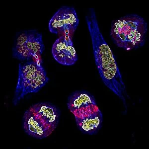
Mitosis. Fluorescence micrograph of two cells during mitosis

![]()
Wall Art and Photo Gifts from Science Photo Library
Mitosis. Fluorescence micrograph of two cells during mitosis
Mitosis. Fluorescence micrograph of two cells during mitosis (nuclear division), when two daughter nuclei are formed from one parent nucleus. The cell on the left is at the prometaphase stage, while the cell on the right is in metaphase. During prometaphase, the nuclear envelope breaks down, allowing the mitotic spindle (red) to interact with the chromosomes (white). At metaphase, the chromosomes line up along the centre of the cell, and the spindles grow from each pole to the chromosomes. Chromosomes are made up of two identical sister chromatids, which are separated into the two daughter nuclei, so that each new cell retains the parent cells genetic information
Science Photo Library features Science and Medical images including photos and illustrations
Media ID 6453997
© DR PAUL ANDREWS, UNIVERSITY OF DUNDEE/ SCIENCE PHOTO LIBRARY
Bipolar Centromere Centromeres Centrosome Chromatid Chromatids Chromosome Chromosomes Cytological Cytology Cytoskeletal Cytoskeleton Dividing Division Fixed Fluorescence Micrograph Fluorescent Hela Cell Metaphase Microscope Microtubule Microtubules Mitosis Mitotic Nuclear Poles Prometaphase Segregating Segregation Separating Spindle Spindles Stained Structures Wide Field Deconvoluted Cells Deoxyribonucleic Acid Genetics
MADE IN THE USA
Safe Shipping with 30 Day Money Back Guarantee
FREE PERSONALISATION*
We are proud to offer a range of customisation features including Personalised Captions, Color Filters and Picture Zoom Tools
SECURE PAYMENTS
We happily accept a wide range of payment options so you can pay for the things you need in the way that is most convenient for you
* Options may vary by product and licensing agreement. Zoomed Pictures can be adjusted in the Cart.