Home > Animals > Insects > Butterflies > Bagworm
Earthworm, transverse section

![]()
Wall Art and Photo Gifts from Science Photo Library
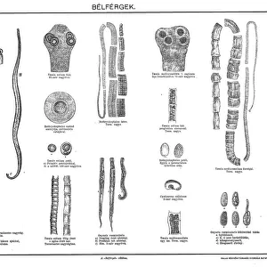
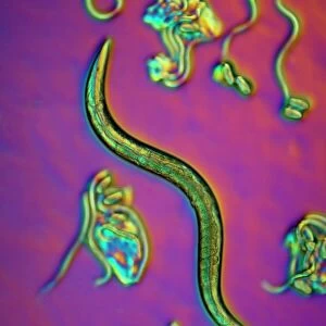
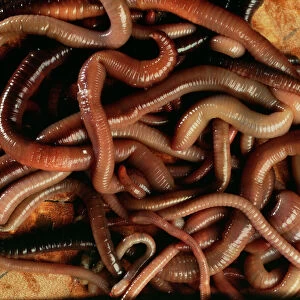
Earthworm, transverse section
Earthworm. Light micrograph of a transverse section through the body of a round segmented earthworm (Lumbricus terrestris) in the intestinal region. The intestine (circular, centre) includes the typhlosole (upper centre). In the body cavity (white) are the excretory organs, the nephridia (kidney-like organs, pink, right and left), and the nerve cord (pink, lower centre). The outer ring (pink) consists of the epidermis, circular muscles, and longitudinal muscle (radiating lines). The gaps in the outer ring are bristles (chaetae) that help the movement of earthworms in their burrows. Magnification: x14 when printed at 10 centimetres across
Science Photo Library features Science and Medical images including photos and illustrations
Media ID 6469003
© DR KEITH WHEELER/SCIENCE PHOTO LIBRARY
Annelid Worm Annelida Bristle Bristles Cross Section Earth Worm Epidermis Histological Histology Intestinal Intestine Longitudinal Muscle Lumbricus Terrestris Transverse Worm Light Micrograph Light Microscope Section Sectioned
FEATURES IN THESE COLLECTIONS
> Animals
> Insects
> Butterflies
> Bagworm
> Animals
> Insects
> Butterflies
> White Cutworm
> Animals
> Worms
> Bristle Worm
> Animals
> Worms
> Segmented Worm
> Arts
> Still life artwork
> Still life art
> Nature-inspired artwork
> Arts
> Still life artwork
> Nature-inspired art
> Arts
> Portraits
> Still life artwork
> Nature-inspired artwork
EDITORS COMMENTS
This print showcases the intricate inner workings of an earthworm, specifically a transverse section through its body. The image reveals the fascinating anatomy and biology of this round segmented creature, known as Lumbricus terrestris. In the center of the image lies the intestine, with its circular shape and prominent typhlosole in the upper center. Surrounding it is the body cavity, where we can observe the excretory organs called nephridia on both sides. These kidney-like structures are depicted in a striking pink color against a white background. The nerve cord is another noteworthy feature captured in this photograph, appearing as a vibrant pink line running along the lower center. It serves as an essential component for transmitting signals throughout an earthworm's body. The outer ring of this cross-section displays various elements crucial to an earthworm's movement and survival. The pink hue represents not only the epidermis but also circular muscles and longitudinal muscle fibers that aid in locomotion. Additionally, we can see gaps within this ring which house bristles called chaetae - these play a vital role in helping earthworms navigate their burrows. Overall, this stunning light micrograph provides us with invaluable insights into Earth's diverse wildlife by showcasing one of nature's remarkable creatures up close and personal.
MADE IN THE USA
Safe Shipping with 30 Day Money Back Guarantee
FREE PERSONALISATION*
We are proud to offer a range of customisation features including Personalised Captions, Color Filters and Picture Zoom Tools
SECURE PAYMENTS
We happily accept a wide range of payment options so you can pay for the things you need in the way that is most convenient for you
* Options may vary by product and licensing agreement. Zoomed Pictures can be adjusted in the Cart.