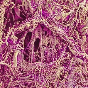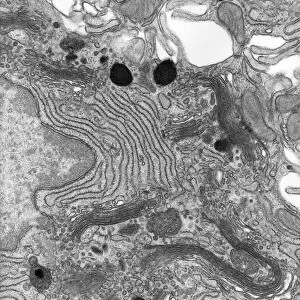
Fallopian tube, SEM

![]()
Wall Art and Photo Gifts from Science Photo Library
Fallopian tube, SEM
Fallopian tube. Coloured scanning electron micrograph (SEM) of a freeze-fractured section through a Fallopian tube (oviduct). The interior (lumen) of the tube (dark space, upper centre) is lined by a thick, highly-folded mucosal layer. A thin muscle layer surrounds this, best seen as the thin compact line at lower centre. These two layers make up the central and upper region of the image. The outer regions are the serosal outer layer of connective tissue (light brown). The two Fallopian tubes are the muscular tubes through which ova (eggs, the female reproductive cells) pass from the ovaries to the uterus following ovulation. Magnification: x100 at 6x7cm size
Science Photo Library features Science and Medical images including photos and illustrations
Media ID 6449955
© STEVE GSCHMEISSNER/SCIENCE PHOTO LIBRARY
Connective Epithelial Lining Epithelium Fallopian Tube Fracture Fractured Freeze Fracture Freeze Fractured Histological Histology Interior Layer Layers Lumen Mucosa Mucosal Mucous Membrane Muscular Oviduct Oviducts Re Production Reproductive System Sections Tissue Tubes Women Section Sectioned Serosa
MADE IN THE USA
Safe Shipping with 30 Day Money Back Guarantee
FREE PERSONALISATION*
We are proud to offer a range of customisation features including Personalised Captions, Color Filters and Picture Zoom Tools
SECURE PAYMENTS
We happily accept a wide range of payment options so you can pay for the things you need in the way that is most convenient for you
* Options may vary by product and licensing agreement. Zoomed Pictures can be adjusted in the Cart.