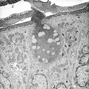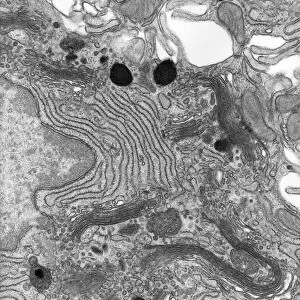
Gastric gland, TEM

![]()
Wall Art and Photo Gifts from Science Photo Library
Gastric gland, TEM
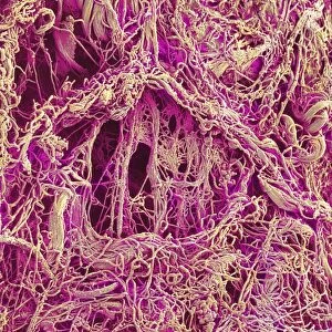
Gastric gland. Transmission electron micrograph (TEM) of a section through the deep region of a gastric (fundus) gland, showing several enzyme-secreting chief cells with many secretory granules, and a single parietal cell, which produces stomach acid. Gastric glands are found in the body, the gastric mucosa, and fundus of the stomach. Chief cells secrete pepsinogen. Acid secretion by the parietal cells converts pepsinogen to pepsin for partial digestion of proteins within the stomach. Both cell types are stimulated by nearby cells, nerves, and local hormones. Magnification: x3000 when printed 10 centimetres tall
Science Photo Library features Science and Medical images including photos and illustrations
Media ID 9242781
© MICROSCAPE/SCIENCE PHOTO LIBRARY
Black And White Bowel Bowels Cell Biology Cytological Cytology Digestion Digestive Digestive System Gastrointestinal Tract Histological Histology Organelle Organelles Pepsin Secretory Stomach Transmission Electron Micrograph Transmission Electron Microscope Cells Fundus Section Sectioned
EDITORS COMMENTS
This print showcases the intricate details of a gastric gland, captured using a transmission electron microscope (TEM). The image reveals the deep region of a gastric (fundus) gland, highlighting several enzyme-secreting chief cells with numerous secretory granules. Additionally, it features a single parietal cell responsible for producing stomach acid. Gastric glands are vital components found in the body's gastric mucosa and fundus region of the stomach. Chief cells play a crucial role by secreting pepsinogen, while parietal cells contribute to acid secretion that converts pepsinogen into pepsin. This conversion aids in the partial digestion of proteins within the stomach. Stimulated by neighboring cells, nerves, and local hormones, both types of cells work harmoniously to support efficient digestion. The magnification level used for this print allows viewers to appreciate these cellular structures at an impressive x3000 when printed 10 centimeters tall. With its monochrome aesthetic and precise focus on cellular biology, this print from Science Photo Library provides valuable insights into the inner workings of our digestive system. It serves as a reminder of how complex yet fascinating our biological processes truly are.
MADE IN THE USA
Safe Shipping with 30 Day Money Back Guarantee
FREE PERSONALISATION*
We are proud to offer a range of customisation features including Personalised Captions, Color Filters and Picture Zoom Tools
SECURE PAYMENTS
We happily accept a wide range of payment options so you can pay for the things you need in the way that is most convenient for you
* Options may vary by product and licensing agreement. Zoomed Pictures can be adjusted in the Cart.