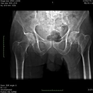
Lung bronchogram, coloured X-ray

![]()
Wall Art and Photo Gifts from Science Photo Library
Lung bronchogram, coloured X-ray
Healthy lung. Coloured bronchogram x-ray of a side (lateral) view of a healthy human lung. The vertebrae of the spinal column are seen at left. At lower right, the bottom two ribs of the rib cage are visible as diagonal bands. From the trachea (not seen) air passes into each lung via a bronchus. This tube branches into many bronchioles (centre, red), to deliver air to every part of the lung. The bronchioles divide to form tiny air sacs called alveoli (not seen) where gaseous exchange occurs. Bronchography renders the airways visible on X-ray film by prior injection of a radio-opaque contrast medium into the windpipe
Science Photo Library features Science and Medical images including photos and illustrations
Media ID 6449637
© PASIEKA/SCIENCE PHOTO LIBRARY
Bronchial Tree Bronchiole Bronchioles Bronchus Diagnosis Diagnostic Technique Lateral Lung Lungs Pulmonary Radiograph Radiography Respiration Respiratory System Spinal Column Vertebrae
EDITORS COMMENTS
This print showcases a vibrant and detailed coloured X-ray of a healthy lung, providing an intricate view of the respiratory system. The lateral perspective allows us to observe the vertebrae of the spinal column on the left side, while at lower right, two diagonal bands represent the bottom ribs of the rib cage. From our unseen trachea, air gracefully enters each lung through bronchi. The bronchus branches out into numerous bronchioles that are prominently displayed in red at the center of this image. These bronchioles play a crucial role in delivering air to every nook and cranny within our lungs. As we delve deeper into this complex network, we discover tiny air sacs called alveoli where essential gaseous exchange occurs; however, they remain unseen in this particular snapshot. Bronchography is responsible for rendering these intricate airways visible on X-ray film by introducing a radio-opaque contrast medium into our windpipe beforehand. This diagnostic technique enables medical professionals to gain valuable insights into normal lung anatomy and diagnose potential abnormalities or diseases affecting respiration. Science Photo Library has expertly captured this remarkable image that not only highlights human body's incredible complexity but also serves as an invaluable resource for scientific research and medical education regarding pulmonary health.
MADE IN THE USA
Safe Shipping with 30 Day Money Back Guarantee
FREE PERSONALISATION*
We are proud to offer a range of customisation features including Personalised Captions, Color Filters and Picture Zoom Tools
SECURE PAYMENTS
We happily accept a wide range of payment options so you can pay for the things you need in the way that is most convenient for you
* Options may vary by product and licensing agreement. Zoomed Pictures can be adjusted in the Cart.